


Overview:
Canine elbow dysplasia is an umbrella term describing several developmental abnormalities that affect the elbow joint and ultimately lead to arthritis.
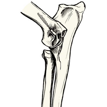
The elbow is a complex joint made up of three bones — the radius, ulna, and humerus — which must align and move together precisely. Any incongruity in their fit or growth leads to uneven force distribution, abnormal wear on the cartilage, and progressive joint damage.
Development of the disease involves many factors but genetics play a major role in the development of elbow dysplasia, which is most commonly seen in large and giant breed dogs.
Normal Elbow
The most common components of elbow dysplasia include:
Joint Incongruity: Abnormal conformation or mismatched growth between the three bones of the elbow at the level of the joint or distantly at the growth plates near the wrist.
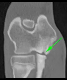
Fragmentation of the Medial Coronoid Process (FMCP): A small bony prominence on the ulna fractures, creating a loose fragment that irritates the joint and accelerates cartilage wear.
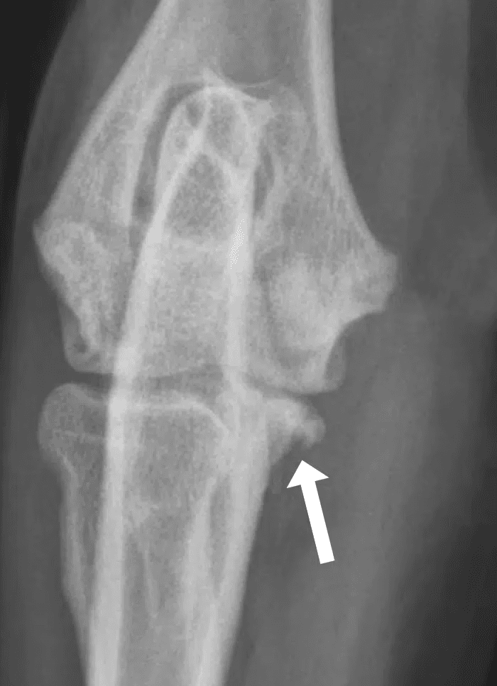
Osteochondritis Dissecans (OCD): A developmental defect in which bone beneath the cartilage fails to form properly, resulting in a weak spot. A cartilage flap can form that causes inflammation and adjacent cartilage wear within the joint.
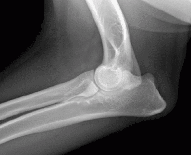
Ununited Anconeal Process (UAP): Failure of a growth plate within the ulna to close normally, leading to separation (fracture) of the anconeal process.
Congenital elbow luxation and incomplete ossification of the humeral condyle (IOHC) are also forms of Elbow Dysplasia but less common.
Joint Incongruity
Short Radius

Signs can begin as early as 4–6 months of age, though most do not show signs until 1-2 years old and many may not have lameness until 5+ years old
Common signs include:
Forelimb lameness that worsens after activity
Stiffness or reluctance to play or exercise
Decreased range of motion in the elbow
Pain or discomfort when the elbow is flexed or extended
Generalized reluctance to run, jump, or bear weight in forelimbs
Diagnosis:
Diagnosis involves a combination of physical examination and imaging. Pain is often evident with manipulation of the elbow.
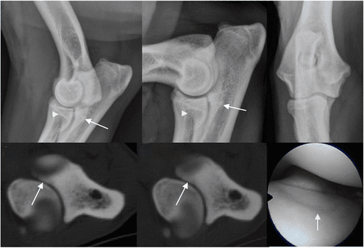
X-rays of the elbow are typically the first diagnostic tool and may reveal fragments, incongruity, or arthritic changes. CT scans or arthroscopy (minimally invasive joint exploration) may be recommended for detailed evaluation, particularly in young patients with mild signs or minimal radiographic changes.
UAP
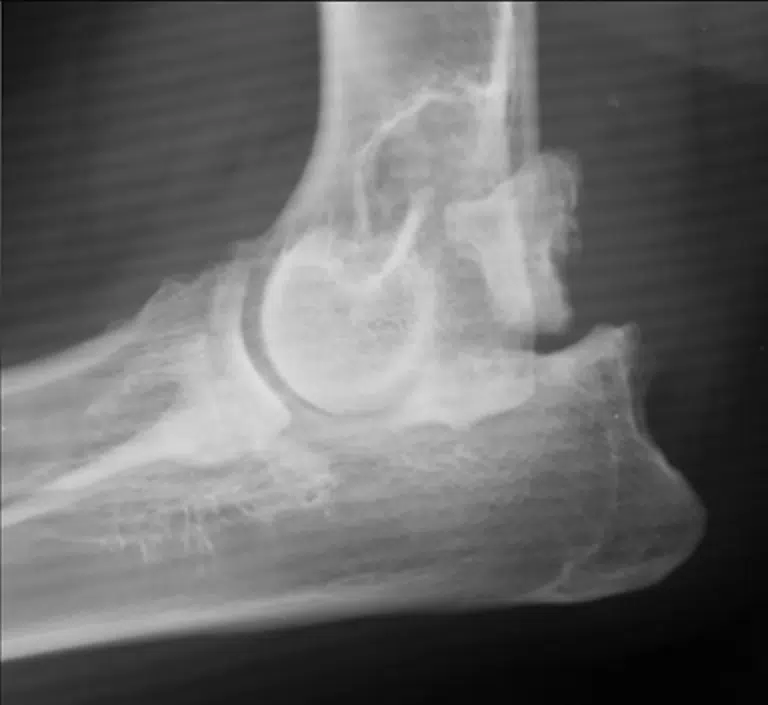
OCD
FMCP
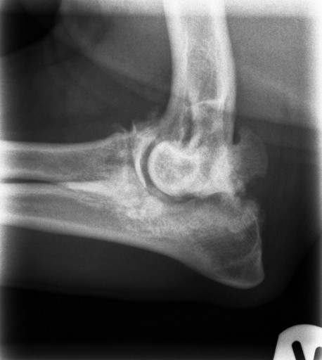
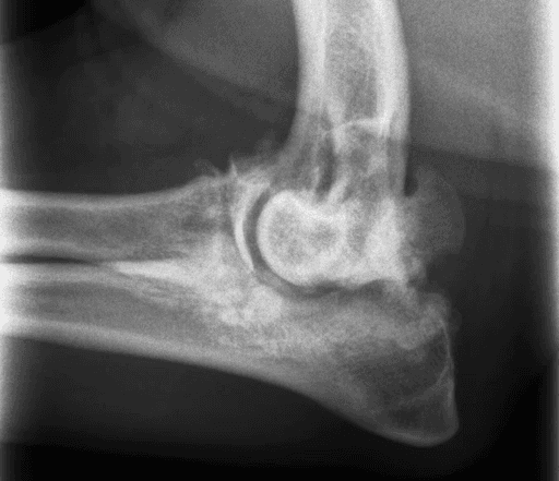
Severe Arthritis
All dogs will see benefit from non-surgical management and some will benefit from surgical intervention.
Conservative management is essential for all dogs with elbow dysplasia, regardless of surgery, to help slow arthritis progression and maintain comfort:
Strict weight management to limit amount of force going through the joints
Consistent Low-impact exercise to maintain a base level of muscle mass
Limit High-impact exercise to limit damage to the elbows and other joints
Rehabilitation therapy to maintain strength and joint mobility
Pain management medications as needed to alleviate pain
Nutraceutical supplementation such as glucosamine, chondroitin, and omega-3 fatty acids to support joint health
Many dogs achieve good long-term comfort and function with consistent conservative management alone.
Surgical Management:
Surgery may be recommended in young dogs to remove painful fragments, restore joint alignment, or slow the progression of arthritis.
Procedures performed at WCVS include:
Joint Incongruity:
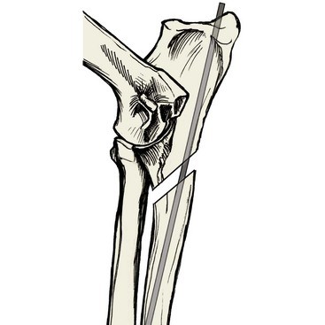
Ulnar Osteotomy/Ostectomy
A cut is made in the ulna to allow the bone to reposition, improving joint congruity.
This procedure is used when the ulna is 'too short' either from irregular growth at the joint surface or when a growth plate injury has desynced growth between the radius and ulna
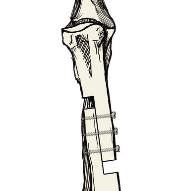
Radial Lengthening
A special series of cuts are made in the radius to allow lengthening of the bone in order to realign the joint surfaces. The bone is then stabilized with a bone plate and screws.
Fragmented Medial Coronoid Process (FMCP):
Arthroscopic Debridement
Arthroscopy is a minimally invasive procedure wherein a small camera and instruments are placed within the joint through very small incisions. The joint is thoroughly inspected for components of elbow dysplasia that were not appreciated on preopative x-rays. The fragmented medial coronoid process is then removed and bed cleaned up of abnormal boney and cartilaginous debris.
Osteochondritis Dissecans (OCD):
Arthroscopic Debridement
Like arthroscopy for FMCP, the joint is inspected through very small incisions. The cartilage flap is removed and the bed cleaned up of abnormal boney and cartilaginous debris.
Ununited Anconeal Process:
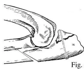
Fragment Fixation
The surface between the fragment and ulna is cleaned up to stimulate healing then the fragment is secured in place with a screw. Generally, an ulnar osteotomy is also performed to protect the screw from breaking due pressure on the anconeal process.
Fragment Removal
The fragment is removed and the bed cleaned of abnormal boney and cartilaginous debris. More arthritis is expected to develop because we do not recreate 'normal' anatomy. However, dogs tend to have improvement of signs and there are no implant complication risks
Postoperative management is dependent on the procedure that was performed but generally involves 6-8 weeks of exercise restrictions.
Outcome is highly dependent on the components present, severity of arthritis already present, age of patient, and adherence to non-surgical management for arthritis long-term. In general, younger dogs with mild disease have a better outcome and see more improvement in their signs. Older dogs with an acute, significant worsening of their signs will likely see improvement. Older dogs with mild or slowly progressive signs may see minimal to no surgical benefit and are likely better treated with non-surgical management alone.
Unfortunately, there is no cure for elbow dysplasia and our surgical options do not prevent the progression of arthritis. Surgical management can help in certain patients but the patients that see the best long-term comfort and functional outcomes are those that consistently adhere to the non-surgical management of arthritis; weight management, activity modifications, rehab, pain medications as needed and supplements.


