


Overview:
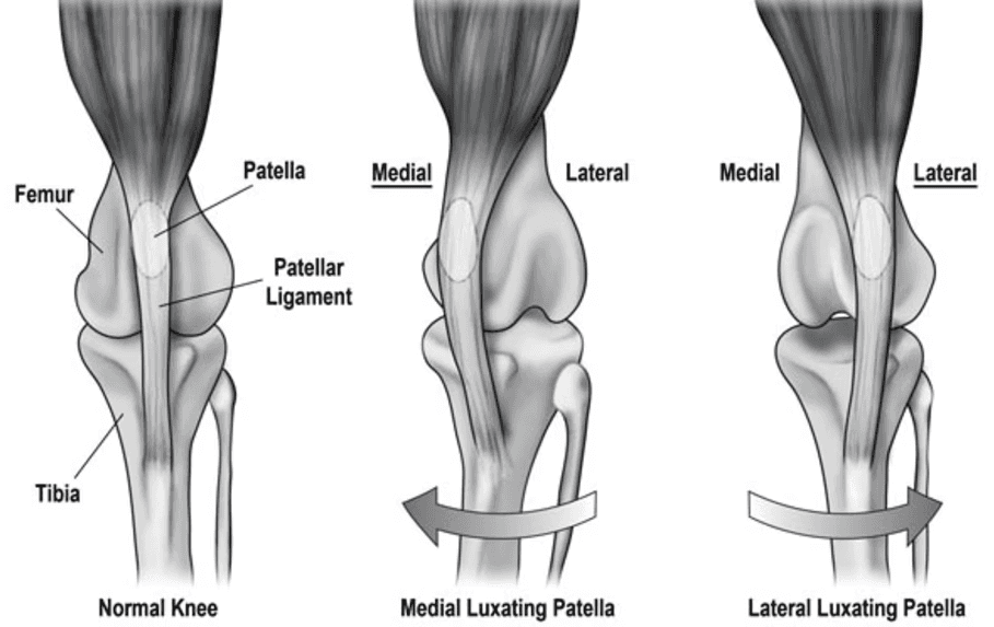
The patella, or kneecap, is a small bone located within the quadriceps muscle that moves up and down within a groove on the femur to help extend the knee.
A patellar luxation occurs when the kneecap dislocates from its normal position and slides to the side of the femur. This can occur towards the inside (medial) or the outside (lateral) of the knee. Medial luxations are most common and can occur in dogs and cats of all sizes while a lateral luxation typically affects large breed dogs with limb deformities
Patellar luxations are graded based on how easily the patella can be displaced and whether it returns to its normal position:
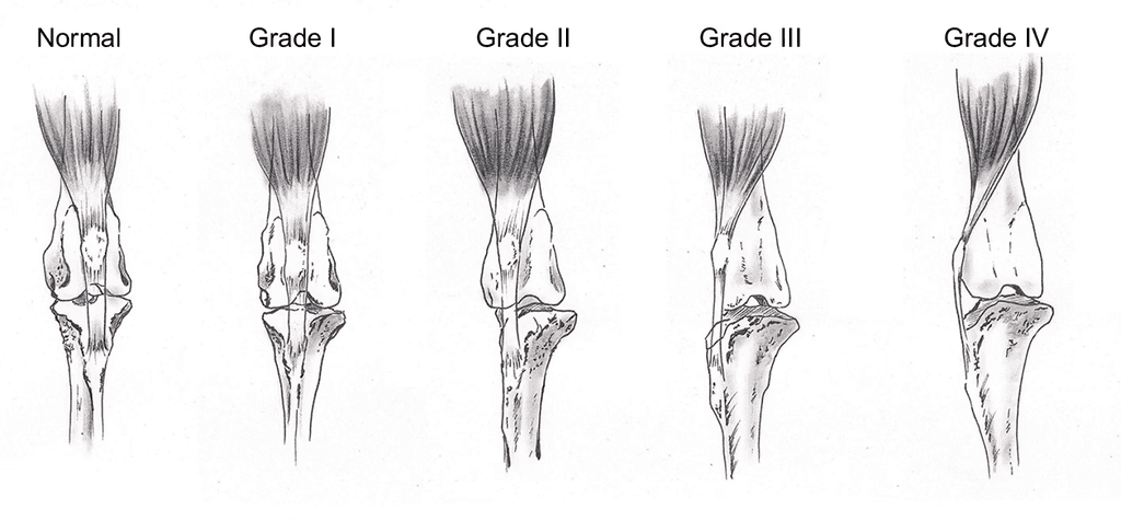
Grade 1: The patella is loose within the groove but does not dislocate completely.
Grade 2: The patella can be dislocated with manipulation but returns to the normal position spontaneously
Grade 3: The patella is dislocated more often than not but it can still be returned to the normal position
Grade 4: The patella remains dislocated and cannot be reduced to the normal position. These are often due to a bone deformity.
Clinical signs will vary but generally correlate with severity of the luxation.
Common signs are:
Intermittent lameness characterized by 'skipping' a few steps but spontaneously returns to normal
Consistent lameness with more severe forms
Stretching or 'mule kicking'
Sitting down or abruptly ceasing activity or play
Resisting or hesitating to jump up on furniture or taking stairs
Over time, repeated luxation events can cause the groove to become shallower and the condition can worsen. Also, dogs and cats with an untreated patella luxation are predisposed to a Cranial Cruciate Ligament Rupture
Diagnosis:
Diagnosis is typically made through a physical exam.
The patella can be luxated during an awake exam in most cases. Thorough examination will include assessment for additional problems (e.g. cranial cruciate rupture, hip dysplasia).
Radiographs are necessary to assess limb alignment and help determine if an additional problem is present.
Patella Luxation
Treatment depends on the severity of the luxation and how it affects your pet’s comfort and mobility. Asymptomatic or mild cases are usually managed conservatively with strict weight control, activity modification, physical therapy, joint supplements, and intermittent pain medications.
Surgical correction is recommended for pets experiencing frequent luxation events, luxation events are affecting quality of life, or in young patients that we want to prevent progression.
Surgical stabilization of the patella luxation is tailored to each patient’s specific needs:
Adjusting soft tissues to balance tension around the knee.
Deepening the femoral groove to better hold the patella in place.
Anti-rotational sutures to control excessive tibial rotation.
Repositioning the tibial tuberosity (where the patellar tendon attaches) to realign the kneecap along the femur
In rare cases, corrective osteotomies are necessary to correct an angular deformity
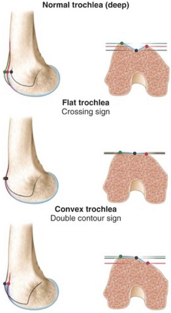
Representation of a shallow trochlear groove
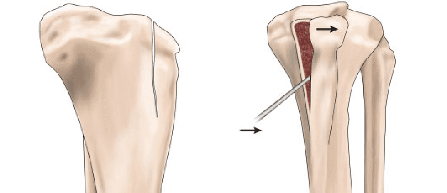
Tibial tuberosity transposition
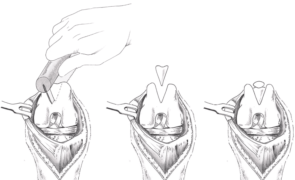
Deepening of the trochlear groove
With proper postoperative care, the success rate for patellar luxation surgery is excellent—typically 90–95%. Recovery generally takes 8–10 weeks and requires very strict activity restriction to protect the repair.
Complications such as incisional infection or implant-associated infection are rare (<3%) when incision care and E-collar use are followed. Implant complications such as pin migration, pin irritation, or implant failure following tibial tuberosity transposition can occur but are uncommon (<5%).
Luxation recurrence rates vary by severity: about <5–10% in grade 2, 15% in grade 3, and 20% in grade 4. When recurrence does occur, it is usually milder and less disruptive to the pet’s comfort.
Large or giant breed dogs, especially those with hip dysplasia or limb deformities, may face a slightly higher complication risk. With appropriate surgical planning, recovery management, and long-term joint care, most pets return to a comfortable, active lifestyle.


